
This is a gem that I've recently dug out of my student practicum notes. It was drawn for me by a very clever sonographer that I worked with for a few months just before graduation.
I'm so glad that I had the chance to work with her. She was a whiz at clarifying the objective of each scan before beginning. And she drilled that lesson into me until I was using her method for every ultrasound.
She taught me that you should always always ALWAYS objectively think about the reason why you are doing each ultrasound. WHY is the patient here for an ultrasound? WHAT are the possible reasons for their symptoms? WHERE would I expect to see pathology? WHAT organs would be involved? HOW will it look? WHAT else needs to be done before the exam is finished?
I know it seems like a lot to think about for each exam, but after a little while it becomes second nature and you'll find yourself doing it all the time. And it will be a huge help in streamlining your exams.
This visual has helped me immensely as a tech for two reasons:
1. I can assess a patient's history and reason for exam and come up with the main objective of the ultrasound. That way I know before I begin what possibilities I am looking for and what is most likely the reason for the patient's symptoms.
2. Using that information I am able to scan with focus and efficiency, I can do more cases both quicker and more thoroughly. Which is not a contradiction, if you do it right.
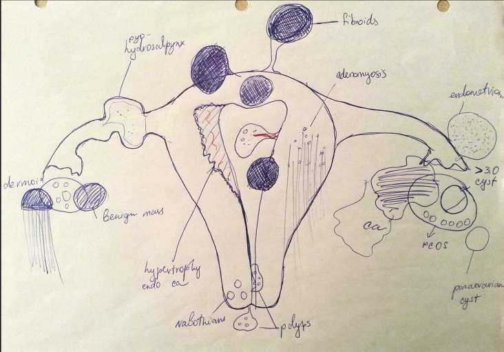
Never forget that, more than knowing how to use a machine and take pictures, we must KNOW what we are looking for, to the best of our ability. We must keep ourselves knowledgeable to best help others. And that starts with having a good idea of what can possibly be found in the pelvis when doing a scan.
I kept this image handy to refer to often and it reminded me of all the little things that I must look for when doing a pelvic exam. It's a great reference and I hope that it comes in handy for you as well.
Happy Scanning!
She taught me that you should always always ALWAYS objectively think about the reason why you are doing each ultrasound. WHY is the patient here for an ultrasound? WHAT are the possible reasons for their symptoms? WHERE would I expect to see pathology? WHAT organs would be involved? HOW will it look? WHAT else needs to be done before the exam is finished?
I know it seems like a lot to think about for each exam, but after a little while it becomes second nature and you'll find yourself doing it all the time. And it will be a huge help in streamlining your exams.
This visual has helped me immensely as a tech for two reasons:
1. I can assess a patient's history and reason for exam and come up with the main objective of the ultrasound. That way I know before I begin what possibilities I am looking for and what is most likely the reason for the patient's symptoms.
2. Using that information I am able to scan with focus and efficiency, I can do more cases both quicker and more thoroughly. Which is not a contradiction, if you do it right.
Never forget that, more than knowing how to use a machine and take pictures, we must KNOW what we are looking for, to the best of our ability. We must keep ourselves knowledgeable to best help others. And that starts with having a good idea of what can possibly be found in the pelvis when doing a scan.
I kept this image handy to refer to often and it reminded me of all the little things that I must look for when doing a pelvic exam. It's a great reference and I hope that it comes in handy for you as well.
Happy Scanning!