
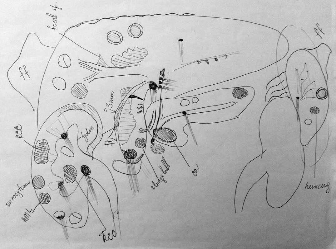
This is the second visual guide that I was given when a student. As you can see, it's a little busier than the pelvic but that's because there's a lot more going on in the upper abdomen.
This is the other great tool that I keep handy to refer to when assessing abdominal organs. If it seems overwhelming at first it helps to focus on just one organ at time.
And believe me, this is about the most complex thing you will every see and it only exists on paper. Let us all take a moment to be grateful for that.
However, this does bring up an interesting and legitimate problem. Complex abdominal pathology does exist and you will eventually come across a scan with multiple pathologies in multiple organs. It can be as a routine check that uncovers a whole lot of unexpected things or as a complex follow-up to multiple other imaging scans. But it happens to us all and the better prepared we are, the easier that scan will be for us.
So what can we do to get through really complex abdominal scans?
1. Be prepared
If you have any previous imaging, reports or numbers, review it to be aware of what you are looking for. If any pathologies need specific follow-up make a note of where they are and what size they were before. If you can, bring a copy of the previous report in the room with you to have handy for reference. Preparation does a lot to make scanning go smoothly.
2. Stay calm
If you have no previous imaging to compare to or no inkling at all that you are about to embark on a challenging scan the best thing to do when you realize what you're in the middle of is to stay calm. The only thing that you need to do is your best and the only way you can do your best is to be relaxed and thinking clearly. Take a few deep breaths, mentally reassess your objective (identify source of RUQ pain or check for an AAA) and use your standard routine to get through everything.
3. One thing at a time
Just focus on one organ at a time. Start with your IVC and Aorta images and move from there through your regular abdominal routine. If you see pathology, assess it, document it, measure it and then move on. Try to approach each organ individually to ensure that it gets your complete attention. You don't want to be distracted by that suspicious looking mass in the kidney and miss dilated ducts in the liver. Each organ needs your full attention so just think about one thing at a time.
4. A review
When you are done assessing and imaging everything in your routine take a moment to mentally or visually review what you have found and figure out if you are forgetting anything. Do a quick check for things like free fluid or abnormal L/Ns or pleural effusions. They are rare but pertinent to complex pathology. It's a final wrap up of imaging that can help give a radiologist a bit more information in a complex case and make you look like a scanning star.
All in all, complex pathology cases are difficult and stressful for most everyone, newbie or experienced. Just strive to keep calm and do your best and you'll always be able to get through them.
Happy Scanning