
I often feel that the cervix doesn't get the attention that it deserves. Sure, it's part of the uterus and we do pay a lot of attention to the uterus, but sometimes it seems that the cervix can get overlooked in the search for fibroids and endometrial thickness.
However, cervical pathology exists and has a stand-alone right to recognition. Not to mention, when you find cervical pathology it makes you look like a superstar ultrasound tech! So here's how I suggest we recognise the cervix.
However, cervical pathology exists and has a stand-alone right to recognition. Not to mention, when you find cervical pathology it makes you look like a superstar ultrasound tech! So here's how I suggest we recognise the cervix.
I have seen situations where cervical pathology was missed due to it being posterior or inferior to the cervix, partially obscured by bowel or bladder wall shadowing or located lower down near the external os.
There are, in fact, so many reasons that cervical pathology can be missed that I've found a dedicated assessment of the cervix was a necessary addition to my scanning routine to prevent overlooking anything.
These are my two main areas of cervical focus:
1. When doing a transabdominal pelvic scan, being sure to take dedicated sagittal and transverse images of the cervix that have been optimised for BEST DETAIL OF THE CERVIX (the cervix usually requires an adjustment in focus and gain different from the uterus for best visualization).
2. When doing an endovaginal pelvic scan the cervix is the first thing that the camera comes into contact with and it's easy to take advantage of that to assess and image the cervical canal before moving on the the uterus as a whole.
Now recently, I came across a case that is a perfect example of tricky cervical pathology:
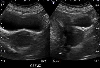
Now, take a look at the image above. Not too shabby considering the empty bladder.
This was a scan done for checkup of IUCD placement. And the IUCD can clearly be seen within the endometrium, no problems there. Objective achieved.
But....
Whats up with that cervix?
It seems a little bulky and that really isn't a nice clear view with the bladder wall shadow falling over it. So let's fiddle around a bit more, push a bit harder, sweep over a bit both left and right and try to get a clearer image...
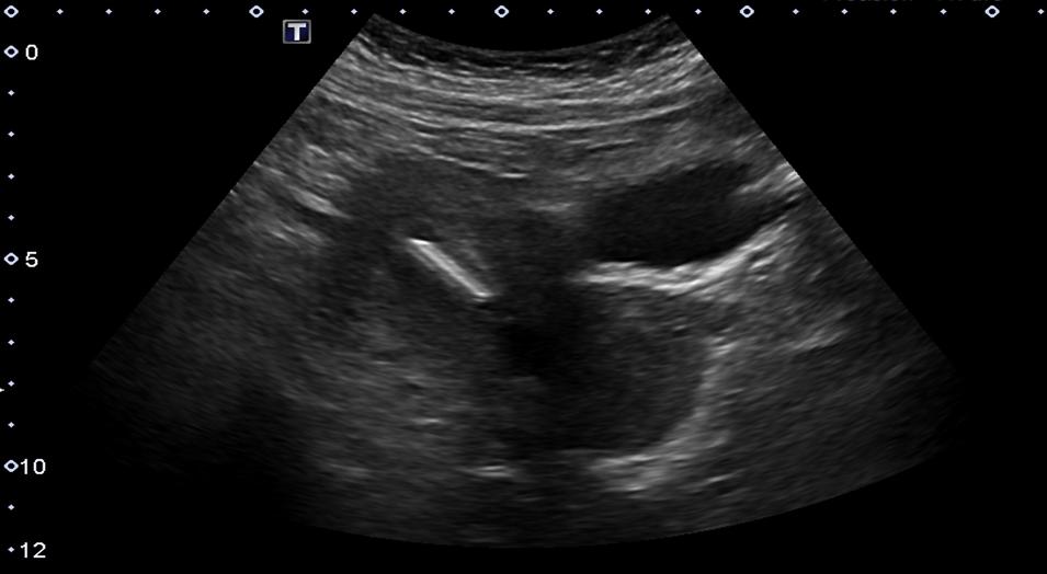
Hmm... a bit clearer of an image and it looks like it the cervix needs further investigation. There could be something at the external os. A fibroid or a mass of some sort.
So let's do an endovaginal scan.
So let's do an endovaginal scan.
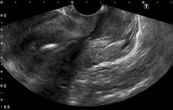
Okay, here we are. There's definitely something in the cervix.
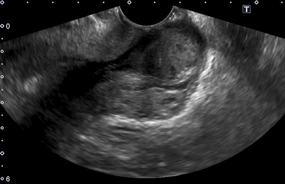
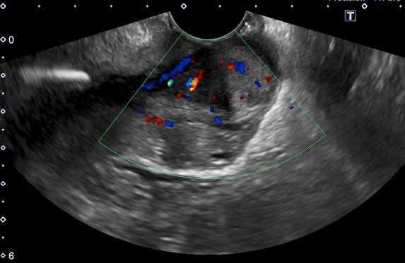
Yup, that's not right. There really was something in the cervical area, and I'm sure glad that I did a focused assessment of the cervix and followed up my uncertainty with further imaging.
Possibly it's just a fibroid but I'm glad to have found this so that the patient can have it followed up.
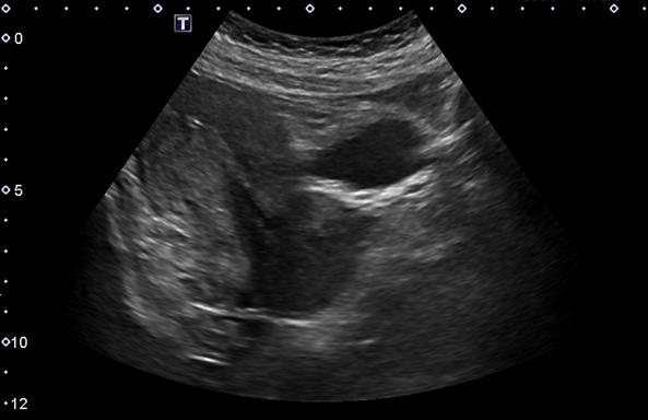
Afterwards I went back to check her history. There had been a previous scan done about 3 years ago, but no mention of a cervical fibroid or mass in the report. However, I notice that there aren't any dedicated cervical images saved, just images of the uterus and cervix together, such as the one below.
Possibly it's just a fibroid but I'm glad to have found this so that the patient can have it followed up.
Afterwards I went back to check her history. There had been a previous scan done about 3 years ago, but no mention of a cervical fibroid or mass in the report. However, I notice that there aren't any dedicated cervical images saved, just images of the uterus and cervix together, such as the one below.
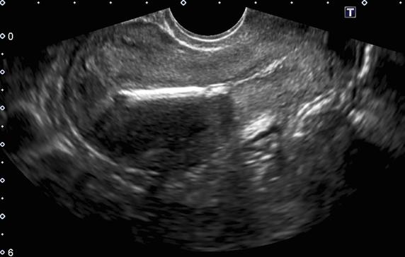
Since the cervix isn't imaged in it's entirety, I can't see clearly, from this picture, all the way from INTERNAL os to EXTERNAL os. So I can't be certain if the mass was not there before, or if it was missed.
This case is a great example of the importance of separate cervical assessment in pelvic scan. And from it I was reminded that if I don't pay attention to an area I will always risk missing something.
So keep a close eye on that cervix and Happy Scanning!
This case is a great example of the importance of separate cervical assessment in pelvic scan. And from it I was reminded that if I don't pay attention to an area I will always risk missing something.
So keep a close eye on that cervix and Happy Scanning!