
We all know about the traditional uses for a high resolution linear transducer; the thyroid, superficial lumps and bumps (why hello there lipoma!), hernias, vascular imaging (the ubiquitous carotids and leg veins) and query appendicitis.
But what about non-traditional uses? As the practice of ultrasound evolves it is benefiting from the creative use of angles, frequency and positions thought up by smart techs. All done in the name of getting that ideal image that helps with a diagnosis. Interested in a little out-of-the-box ultrasound thinking? Here it is…
But what about non-traditional uses? As the practice of ultrasound evolves it is benefiting from the creative use of angles, frequency and positions thought up by smart techs. All done in the name of getting that ideal image that helps with a diagnosis. Interested in a little out-of-the-box ultrasound thinking? Here it is…
You’ll Be Surprised By What You See…
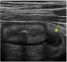
1. CROWN RUMP LENGTH
Want to sharpen up the edges of that teeny tiny baby bean? A linear transducer might be just the thing to help.
A 7-9 MHz linear transducer is commonly used in the 11-13 week NT Scan to assess the nuchal thickness of a fetus, and it can also help out on a routine OB under scan to provide a clearer image for measurement of the CRL or to assess for pathology.
A 7-9 MHz linear transducer is commonly used in the 11-13 week NT Scan to assess the nuchal thickness of a fetus, and it can also help out on a routine OB under scan to provide a clearer image for measurement of the CRL or to assess for pathology.
2. Bowel
Think back to all those LLQ pain histories on your requisitions.
What is predominantly in the LLQ? Some lovely large intestine, that’s what. And a pathology commonly affecting the colon in that area is diverticulitis, the inflammation of bowel diverticulum (by the way, let’s all eat more fiber to help prevent this!) which can be imaged with high frequency linear ultrasound.
If you are not familiar with what an inflamed diverticulum looks like check out ultrasoundcases.info in their Abdomen Folder for teaching cases and examples. It’s pretty neat to see and you’ll be helping someone out who may need treatment for this affliction.
3. Adventurous Ovaries
Speaking of the LLQ, how about ovary assessment? Some ovaries start a migration out of the lesser pelvis and into the greater pelvis, otherwise known as the lower quadrants of the abdomen.
Up there they often sit quite superficially among the bowel and are more clearly imaged with a linear transducer instead of a curved one. So the next time you have trouble finding a little ovary down in the pelvis try looking up a little higher, you might be surprised by what you can find up there.
Up there they often sit quite superficially among the bowel and are more clearly imaged with a linear transducer instead of a curved one. So the next time you have trouble finding a little ovary down in the pelvis try looking up a little higher, you might be surprised by what you can find up there.
4. Pancreas
Yes, this will really only work on slender, fairly gasless patients, but they do exist and they do come for ultrasounds. And if you do happen to be scanning one you’ll often find that a linear transducer gives a much clearer image of the pancreas than the curvy. This is important when assessing for pancreatitis, pancreatic CA and peri-pancreatic lymph nodes. And you’ll look like a star tech!
5. Gallbladder
Ever notice how that superficial fundal portion of the gallbladder can be obscured by artifact when using a curvy transducer? That’s worrisome when assessing for polyps or wall thickening. You’ll be able to much better assess that portion of a gallbladder with a linear transducer and have some very pretty pictures to show for it. Give it a try next time.
There is no end to the possibilities seen with a linear transducer and as the technology improves, there will only be more and more things to be seen. So jump in and try something new, you have nothing to lose and a whole lot of lovely images to gain.
Happy Scanning!
Happy Scanning!